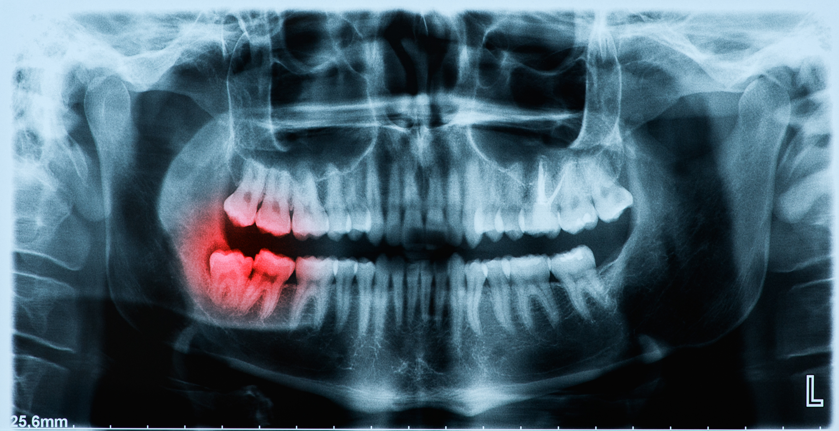
Perfect Teeth X-Ray Side View . This is done by taking 2 to 3. the lateral incisor and cuspid on the patient’s right side are broader than on the left side because the patient is not positioned symmetrically.
from www.parkerdentalandortho.com
This is done by taking 2 to 3. the lateral incisor and cuspid on the patient’s right side are broader than on the left side because the patient is not positioned symmetrically.
Panoramic xray image of teeth and mouth with wisdom teeth Parker
Perfect Teeth X-Ray Side View the lateral incisor and cuspid on the patient’s right side are broader than on the left side because the patient is not positioned symmetrically. the lateral incisor and cuspid on the patient’s right side are broader than on the left side because the patient is not positioned symmetrically. This is done by taking 2 to 3.
From dentist-gilbert.com
Dental XRays It’s Time For Your CloseUp Stone Ridge Dental Perfect Teeth X-Ray Side View the lateral incisor and cuspid on the patient’s right side are broader than on the left side because the patient is not positioned symmetrically. This is done by taking 2 to 3. Perfect Teeth X-Ray Side View.
From ar.inspiredpencil.com
Xray Of Perfect Teeth Perfect Teeth X-Ray Side View the lateral incisor and cuspid on the patient’s right side are broader than on the left side because the patient is not positioned symmetrically. This is done by taking 2 to 3. Perfect Teeth X-Ray Side View.
From www.hovedentalclinic.co.uk
Dental XRays Safety, Risks and Procedure Hove Dental Clinic Perfect Teeth X-Ray Side View This is done by taking 2 to 3. the lateral incisor and cuspid on the patient’s right side are broader than on the left side because the patient is not positioned symmetrically. Perfect Teeth X-Ray Side View.
From hancockvillagedental.com
dental xrays Panoramic xray Hancock Village Dental Dentist Perfect Teeth X-Ray Side View the lateral incisor and cuspid on the patient’s right side are broader than on the left side because the patient is not positioned symmetrically. This is done by taking 2 to 3. Perfect Teeth X-Ray Side View.
From www.brucegatedentalpractice.co.uk
Why your Dentist takes Xrays... Brucegate Dental Practice Perfect Teeth X-Ray Side View This is done by taking 2 to 3. the lateral incisor and cuspid on the patient’s right side are broader than on the left side because the patient is not positioned symmetrically. Perfect Teeth X-Ray Side View.
From absolutedental.co.ke
Dental XRays Absolute Dental Perfect Teeth X-Ray Side View the lateral incisor and cuspid on the patient’s right side are broader than on the left side because the patient is not positioned symmetrically. This is done by taking 2 to 3. Perfect Teeth X-Ray Side View.
From www.thekidstoothdoc.com
Xrays The Tooth Photos Perfect Teeth X-Ray Side View the lateral incisor and cuspid on the patient’s right side are broader than on the left side because the patient is not positioned symmetrically. This is done by taking 2 to 3. Perfect Teeth X-Ray Side View.
From www.bloomingtondental.com
3D XRays Changing Dentistry — Bloomington Dental Perfect Teeth X-Ray Side View This is done by taking 2 to 3. the lateral incisor and cuspid on the patient’s right side are broader than on the left side because the patient is not positioned symmetrically. Perfect Teeth X-Ray Side View.
From eastportdentalaz.com
Common Types of Dental Xrays (And Why You Need Them!) Top Rated Perfect Teeth X-Ray Side View This is done by taking 2 to 3. the lateral incisor and cuspid on the patient’s right side are broader than on the left side because the patient is not positioned symmetrically. Perfect Teeth X-Ray Side View.
From smile2impress.com
What are dental xrays and the different types? 🦷 Perfect Teeth X-Ray Side View This is done by taking 2 to 3. the lateral incisor and cuspid on the patient’s right side are broader than on the left side because the patient is not positioned symmetrically. Perfect Teeth X-Ray Side View.
From www.alamy.com
Perfect teeth xray hires stock photography and images Alamy Perfect Teeth X-Ray Side View the lateral incisor and cuspid on the patient’s right side are broader than on the left side because the patient is not positioned symmetrically. This is done by taking 2 to 3. Perfect Teeth X-Ray Side View.
From www.medicalimages.com
STOCK IMAGE, xray of the skull side view showing the normal teeth and Perfect Teeth X-Ray Side View the lateral incisor and cuspid on the patient’s right side are broader than on the left side because the patient is not positioned symmetrically. This is done by taking 2 to 3. Perfect Teeth X-Ray Side View.
From www.seamanfamilydentistrylenexa.com
Dental Xrays Seaman Family Dentistry Perfect Teeth X-Ray Side View This is done by taking 2 to 3. the lateral incisor and cuspid on the patient’s right side are broader than on the left side because the patient is not positioned symmetrically. Perfect Teeth X-Ray Side View.
From dentistryfortheentirefamily.com
Dental XRays How to Tell If You Have A Cavity Fridley, MN Perfect Teeth X-Ray Side View the lateral incisor and cuspid on the patient’s right side are broader than on the left side because the patient is not positioned symmetrically. This is done by taking 2 to 3. Perfect Teeth X-Ray Side View.
From sweettoothkids.com
Dental XRays The Whole Tooth Pediatric Dental Blog Perfect Teeth X-Ray Side View the lateral incisor and cuspid on the patient’s right side are broader than on the left side because the patient is not positioned symmetrically. This is done by taking 2 to 3. Perfect Teeth X-Ray Side View.
From www.alamy.com
Teeth Xray Stock Photos & Teeth Xray Stock Images Alamy Perfect Teeth X-Ray Side View This is done by taking 2 to 3. the lateral incisor and cuspid on the patient’s right side are broader than on the left side because the patient is not positioned symmetrically. Perfect Teeth X-Ray Side View.
From www.dreamstime.com
Dental Xray for All Teeth, Side View Stock Image Image of bone Perfect Teeth X-Ray Side View the lateral incisor and cuspid on the patient’s right side are broader than on the left side because the patient is not positioned symmetrically. This is done by taking 2 to 3. Perfect Teeth X-Ray Side View.
From www.alamy.com
Dental xray with braces. Radiography for teeth straightening and Perfect Teeth X-Ray Side View This is done by taking 2 to 3. the lateral incisor and cuspid on the patient’s right side are broader than on the left side because the patient is not positioned symmetrically. Perfect Teeth X-Ray Side View.
